
Movement Assessment Versus Imaging: What Wins?
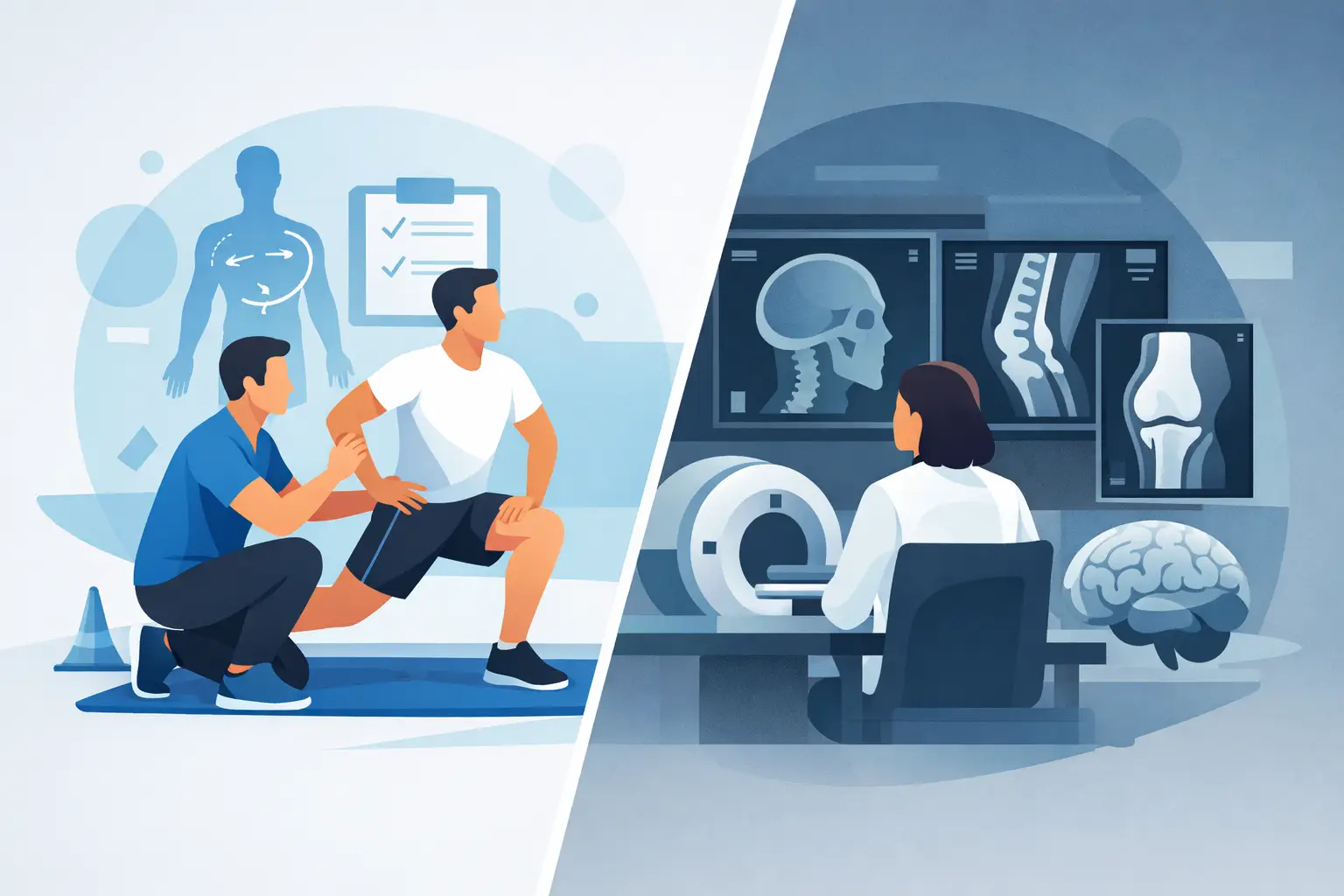
A shoulder MRI can show a tear. A lumbar X-ray can show arthritic changes. But neither test can tell you why your knee caves in when you land, why your back tightens after nine holes of golf, or why pain returns every time you increase your running mileage. That is where movement assessment versus imaging becomes a meaningful decision, not just a medical one.
For active adults and athletes, the goal is rarely to collect more information for its own sake. The goal is to understand what is limiting you, build a clear plan, and return to training, work, sport, and daily life with confidence. Imaging can be valuable. Movement assessment can be essential. The right choice depends on the question that needs to be answered.
Movement Assessment Versus Imaging: Different Tools, Different Questions
Imaging looks at structure. X-rays, MRIs, CT scans, and diagnostic ultrasound can reveal bones, joints, tendons, ligaments, discs, and other tissues. When a clinician needs to rule out a fracture, identify a complete tendon rupture, assess a significant traumatic injury, or investigate symptoms that may require medical intervention, imaging can provide critical information.
A movement assessment looks at function. It examines how your body performs the tasks that matter to you: walking, squatting, hinging, reaching, rotating, running, lifting, landing, or changing direction. It also considers mobility, strength, coordination, balance, joint control, training history, and symptom behavior.
Neither approach replaces the other. The mistake is assuming that a picture of a body part automatically explains a movement problem.
A scan may identify a rotator cuff tear in someone with shoulder pain. Yet two people with the same finding can have very different experiences. One may lift overhead without meaningful limitation. The other may hurt during pressing, lack shoulder control, compensate through the neck, and avoid the gym. The structural finding matters, but the way that person moves, loads the tissue, and responds to activity matters too.
Why Imaging Findings Do Not Always Equal the Source of Pain
Modern imaging is highly sensitive. That is useful when a serious injury is suspected, but it also means scans often show age-related or previously existing changes. Disc bulges, meniscus changes, tendon wear, and mild joint degeneration can appear in people who have no pain at all.
That does not mean imaging is wrong. It means imaging needs context. A finding on a report must be matched with your symptoms, physical examination, injury mechanism, and functional limitations before it is treated as the cause.
Consider a runner with hip pain. An image may show mild labral changes, but the more actionable issue may be limited hip extension, poor single-leg control, a sudden spike in weekly mileage, or inadequate strength through the glutes and trunk. If the plan focuses only on the report, the runner may never address the factors that made running painful in the first place.
This is also why people can feel discouraged after receiving imaging results that sound alarming. A phrase such as degeneration or tear can create the impression that movement is dangerous. In many cases, the more productive question is not simply, What does the scan show? It is, What can you do now, what reproduces symptoms, and what needs to improve for you to return to the activities you value?
What a Quality Movement Assessment Reveals
A quality assessment is more than watching someone do a few stretches. It begins with a detailed conversation about the problem: when it started, how it changed, what aggravates it, what eases it, what training looked like before symptoms began, and what outcome the person wants.
From there, the clinician evaluates the movements connected to the complaint. For a golfer with low back pain, that may include hip rotation, thoracic mobility, hinging mechanics, single-leg control, trunk strength, and elements of the golf swing. For a tennis player with elbow pain, it may include shoulder strength, grip tolerance, wrist control, serving volume, and how force is transferred through the body.
The result should be specific. Rather than telling you that you are tight or weak, a strong assessment identifies relevant limitations and explains how they connect to your goal. It also creates measurable starting points. Can you squat to depth? Run for 20 minutes? Carry groceries without pain? Perform a controlled single-leg step-down? Those benchmarks help guide progression and show whether the plan is working.
At Back In Motion, this root-cause approach is central to The Gray Method™. The process is designed to connect symptoms with movement limitations, then progress clients from pain relief and mobility work into strength, capacity, and performance. That matters because feeling better for a week is not the same as being prepared for the demands of your sport or lifestyle.
When Imaging Is the Right Next Step
There are times when imaging should move higher on the priority list. A major traumatic injury, visible deformity, inability to bear weight after an injury, suspected fracture, sudden significant weakness, or a joint that feels unstable after a collision may warrant prompt medical evaluation and imaging.
Certain symptoms also require urgent attention. These can include loss of bowel or bladder control, numbness in the saddle region, rapidly worsening weakness, unexplained fever with severe pain, unexplained weight loss, or pain associated with a history that raises concern for serious disease. In these situations, do not try to train through the problem or rely on a general exercise plan. Seek appropriate medical care.
Imaging can also be helpful when progress does not match expectations. If a well-executed rehab plan is not improving symptoms, if pain persists despite appropriate load management, or if examination findings suggest a more significant tissue injury, imaging may help clarify the next decision. The point is not to avoid scans. It is to order and interpret them for a clear clinical reason.
When Movement Assessment Should Lead
For many non-traumatic aches, recurring injuries, training-related pain, and performance plateaus, movement assessment is often the more useful starting point. This is especially true when you can still move but do not trust the movement, when pain appears only during certain tasks, or when you have already had imaging and still do not know what to do next.
A person with recurring back pain may not need another scan to start improving their hip mobility, trunk endurance, lifting strategy, and tolerance to progressively heavier loads. A runner with shin pain may need a closer look at training volume, calf strength, ankle motion, footwear changes, and running mechanics. A new parent dealing with pelvic discomfort may need an individualized pelvic health assessment that considers pressure management, breathing, strength, and daily demands rather than a generic core routine.
The practical advantage is immediate direction. A movement assessment can identify what to modify today, what to strengthen this week, and what criteria should be met before returning to higher-demand activity.
The Best Plan Uses Both Information and Progression
The most effective care does not create a false competition between movement assessment and imaging. It uses the right information at the right time.
If imaging confirms a tendon injury, the next question is still functional: how much load can the tendon tolerate, what movement patterns need attention, and how will strength be rebuilt? If a scan shows common age-related changes, the next question is still functional: which movements are limited, what symptoms are modifiable, and how can confidence be restored?
This is where generic treatment often falls short. Passive care may temporarily reduce symptoms, but it does not automatically prepare you to deadlift, sprint, serve, pick up your child, or play 18 holes. Long-term results require progressive exposure to the demands you want to handle.
A clear plan should evolve as you improve. Early sessions may focus on reducing irritation and restoring comfortable motion. The next phase should build strength, control, and capacity. The final phase should resemble real life or sport, whether that means decelerating on one leg, carrying heavy loads, rotating under control, or tolerating repeated overhead work.
The best assessment is the one that leads to better decisions. If you have a scan, use it as one piece of the picture. If you do not have a scan, do not assume you are missing the answer. Your body is not only a collection of structures. It is a system built to move, adapt, and get stronger when the plan matches the problem.
About the Author: Dr. Scott Gray





 Call (239) 788-0838